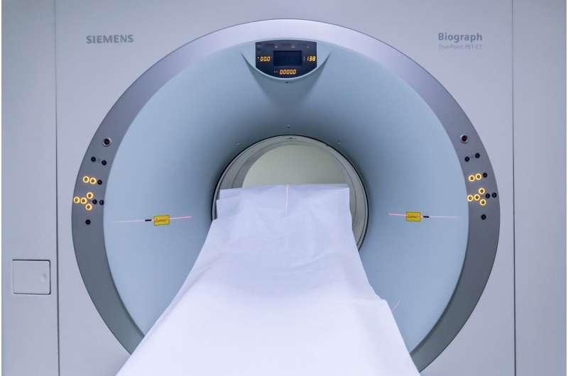
Computed tomography (CT) scanning, which creates detailed 3D images of bones, soft tissues, and organs, is better than standard X-rays for investigating complex injuries, cancers, and vascular issues.
The next generation of CT scanners is starting to use a new technology called photon-counting computed tomography (PCCT), at the heart of which are special detectors that count individual X-ray photons and measure their energy. The sharper, higher-resolution images the new machines produce means better visualization of fine structures (e.g., bronchi) and tissue composition—for more accurate—and possibly earlier—diagnosis in fields such as cardiology, neurology, and cancer screening.
Researchers from the University of Victoria and a leading BC-based provider of X-ray imaging technology are using the Canadian Light Source at the University of Saskatchewan to find ways to improve the ultrathin metal layer (contact) on the top of the detector, which plays a critical role in its performance. Their paper is published in the Journal of Materials Science: Materials in Electronics.
“Any time you give medical professionals more detailed, accurate information about your body, it’s useful for diagnosis and better outcomes,” said Dr. Tom Tiedje, professor emeritus in electrical engineering at the University of Victoria and the University of British Columbia.
Using the SGM beamline at the CLS, the researchers discovered that the contact material currently used (an oxide)—which is only a few atoms thick—”is kind of a complicated beast.”: It controls the extraction of electrons from the detector. Tiedje and his colleagues are now assessing the performance of an alternative material (a sulfide).
“I feel like we’re making progress,” he says. “I think there’s good potential for improving the device performance.”
Better contacts on X-ray detectors could mean CT scanners will be able to do an even better job of differentiating between body tissues of different densities. “X-rays have different wavelengths,” explained Tiedje, “and body tissues respond differently to X-rays.” Current scanners can distinguish, for example, bone from muscle but not muscle from blood vessels. Today’s scanners measure how bright the X-ray photons are as they pass through tissue.
Next-gen detectors will count the photons “one by one, billions and billions of them,” and allow colors to be assigned to different wavelengths that can then distinguish different tissues of similar densities.
The result is a more detailed picture. There’s still more work to be done, says Tiedje. “But if you can detect a small thing like a growth that looks the same as surrounding tissue, you won’t have to wait until it gets big to see it on a CT scan, so that’s definitely an advantage.”
More information
Hao Yuan et al, Composition of oxide on CdZnTe surface and changes caused by X-ray exposure, Journal of Materials Science: Materials in Electronics (2025). DOI: 10.1007/s10854-025-16076-0
Key medical concepts
Clinical categories
The content is provided for information purposes only.