Study using Cubresa SPECT scanner finds potential non-invasive diagnosis for Alzheimer’s
Cubresa Inc., a medical imaging company that develops and markets molecular imaging systems, today announced that their compact SPECT (Single-Photon Emission Computed Tomography) scanner was used in a study by Dalhousie University and Mount Saint Vincent University researchers in Halifax, Nova Scotia to help evaluate the diagnostic potential of a new molecular label that could lead to the early detection of Alzheimer’s disease in living patients.
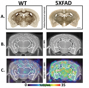
Representative WT (wild-type, left column) and 5XFAD (Alzheimer’s model, right column) brains at the mid-coronal level. Row A: Histochemical staining of BChE, revealing marked elevation of BChE in AD, notably in the cerebral cortex. C = cerebral cortex, H = hippocampus, BG = basal ganglia, Th = thalamus, A = amygdala. Scale bar = 1mm. Row B: CT with co-registered MR at the same level as Row A. Row C: SPECT images acquired at 4 mins post-injection with co-registered CT/MR and ROIs. Marked retention in the cerebral cortex is evident in the 5XFAD brain compared to the WT brain and to a lesser extent in amygdala, hippocampus, basal ganglia and thalamus. Images courtesy of the Darvesh group and BIOTIC.
Alzheimer’s disease (AD) is a progressive neurological condition that causes dementia and for which there is no FDA-approved treatment for the underlying disease. There is a need for a non-invasive way to detect and diagnose the disease in living patients because definitive diagnosis still requires post mortem examination.
The study was performed by Dr. Sultan Darvesh and his research group at BIOTIC (Biomedical Translational Imaging Centre) at the Izaak Walton Killam Health Centre in Halifax, and was partially funded by TREVENTIS™ Corporation of which Dr. Darvesh is a scientific co-founder.
SPECT imaging provides 3D information with a high degree of sensitivity and specificity for detecting the presence of a particular radiolabeled enzyme, in this case the BChE (butyrylcholinesterase) enzyme. Prior research has shown that elevated BChE expression levels are associated with abnormal ß-amyloid (Aß) plaques in the brains of Alzheimer’s patients.
The novel molecular label, TRV6001, was synthesized and determined to bind to BChE with high specificity before being injected into 5XFAD mouse models of Alzheimer’s Disease. TRV6001 is still under development and has not yet been shown to be safe or effective in humans.
“SPECT imaging has provided us the first direct in vivo evidence that the TRV6001 label can cross the blood-brain barrier and actually exhibits higher retention in the brains of AD mice (whose BChE level is markedly elevated) than in normal control brains,” said lead author Drew DeBay, Ph.D. candidate at the Department of Medical Neuroscience, Dalhousie University.
In addition, the retention in BChE-associated brain structures was shown to closely follow the known histochemical distribution of BChE that has been previously established in AD mouse models.
Up to now there have been many false-positives. Previous labels were able to bind to Aß plaque–but Aß is also present in cognitively normal people. BChE, which we are detecting, is notably absent in the non-Alzheimer’s brain.”
Dr. Sultan Darvesh, FRCPC, Professor at the Department of Medical Neuroscience, Dalhousie University.
“Alzheimer’s disease affects so many people and causes such pain and suffering for individuals and their families,” said George Abe, CEO of Cubresa. “We’re pleased to know that our SPECT scanner has contributed to a big step towards developing effective treatments and monitoring their effect.”
The research was presented this year at the Alzheimer’s Association International Conference® (AAIC®), the world’s largest forum for the dementia research community, in Toronto, Canada.
Cubresa